CARDIOMYOPATHY is as the name implies, i.e., pathology of the myocardium, (heart muscle), of the cardia (heart). Initially, it was applied to unknown causes of deteriorating heart muscle function, principally in young, otherwise healthy persons, although, it may also include the old as well. As our knowledge of causes and pathology has improved, there has been a blurring of the distinction between heart muscle malfunction of unknown causes and those of known causes. This classification, exclusively, still focuses on heart muscle abnormalities, in contradistinction to those maladies involving the heart valves, or the covering over the heart (pericardium).
The best classification has revolved around distinct functional or hemodynamic properties. Since most Cardiomyopathies result in Congestive Heart Failure and/or arrhythmias, this classification is of great utility in the treatment of these conditions. Five major forms are recognized: (i) dilated cardiomyopathy, (ii) hypertrophy cardiomyopathy,(iii) restrictive cardiomyopathy, (iv) right ventricular cardiomyopathy and (v) non-classifiable cardiomyopathies with distinct hemodynamic properties. Some of the specific dysfunctions of the heart muscle may overlap in this classification, but the clinical usefulness is still helpful. These functions are expressed as 1.)Dilatation (ventricular enlargement), 2.) Thickness of the heart muscle (Hypertrophy), or 3.) Stiffness of the heart muscle.
Known causes of heart muscle dysfunction, often referred to as Secondary Cardiomyopathies, are also included in categories of Cardiomyopathies. The new World Health Organization’s, World Health Foundation’s (WHO/WHF) definition comprises Inflammatory cardiomyopathy, defined as MYOCARDITIS in association with cardiac muscle dysfunction. Also, Autoimmune, and Infectious forms of cardiomyopathy are recognized. Viral Cardiomyopathy, which may be accompanied by inflammation, has been recognized in some instances. Some cardiomyopathies have a relationship with other organ systems, while still others are hereditary.
The end result of all these different forms of Cardiomyopathies is the worsening of cardiac work. This results, either, from failure of the pump function or from failure of the heart muscle to relax, called ventricular compliance. Hence, the clinical presentation of these Cardiopathies as Congestive Heart Failure. This muscle damage may also interfere with the electrical properties of the heart and result in severe Arrhythmias.
FUNCTIONAL IMPAIRMENT
DILATED CARDIOMYPOATHY
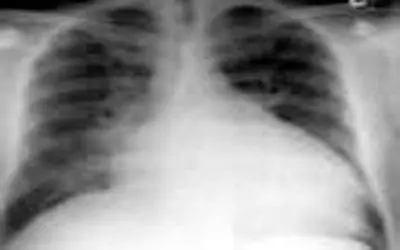
This is a syndrome characterized by a large dilated heart, often accompanied by Congestive Heart Failure. It is frequently referred to as “Congestive Cardiomyopathy”. This is because a dilated heart with stretched cardiac muscle acts like a rubber band, which has been stretched too far and therefore lacks the power to contract. This results in a flabby heart unable to propel the blood forward, i.e., pump failure. The muscle thickness may be normal, increased, or decreased. Most often, the cause of Congested Dilated Cardiomyopathy is elusive and unknown, but it is probably the end result of many forms of heart muscle damage caused by a variety of toxic, metabolic, or infectious agents. Alcohol, pregnancy, hypertension, are a few of the known causes of his syndrome. Once these are ruled out, as well as infection, the syndrome is often referred to as Idiopathic Congestive Cardiomyopathy.
The pathology demonstrates enlargement and dilatation of all four chambers of the heart. The ventricles are more dilated than the atria. Histological examination reveals interstitial and peri- vascular fibrosis in the walls of the ventricles.
The symptoms are those of left ventricular failure, i.e., congestive heart failure. Dyspnea on exertion, orthopnea, paroxysmal nocturnal dyspnea, and rest dyspnea occur. Fatigue and weakness due to diminished cardiac output are prominent. Physical examination reveals a weak pulse, gallop heart rhythm and in the late stages, swollen feet and a swollen liver, associated with abdominal swelling (Ascites).
Treatment, as the syndrome suggests, is directed in strengthening the heart muscles, diminishing the fluid backup, and relieving the heart muscle of resistance to pump against. So Digitalis, which strengthens the heart muscle; diuretics to promote fluid mobilization, and peripheral vasodilators to decrease peripheral resistance are of great help. Other more recent medications, which facilitate the same goals are being incorporated in the treatment with fair results.
Alcoholic cardiomyopathy is probably the most common cause of congestive dilative cardiomyopathy in the western world.
HYPERTROPHIC CARDIOMYOPATHY
This is a condition where the left ventricular wall is thickened (hypertrophied), and the cavity is perforce small. Hypertension is the most common cause of this form of Cardiomyopathy, but an inheritable form, (IHSS-Idiopathic hypertrophy sub-aortic stenosis) is the originator of this classification. Actually, the wall of the left ventricle may be symmetrically hypertrophied, or the hypertrophy may involve the sub aortic, valvular, or supravalvular areas of the ventricle, or only the septum may be asymmetrically involved (ASH).
The pathological examination reveals a marked increase in myocardial mass, with the ventricular cavities being small. The atria are also often hypertrophied and dilated. Myocardial fiber disarray is seen in the interventricular septum in the IHSS cases.
The clinical symptoms include dyspnea from impaired ventricular filling do to the small cavities and the rapid buildup of pressure do the thickened wall stiffness. Fatigue and syncope, as well as angina pectoris are common. Understandably, exertion tends to exacerbate the symptoms. The physical examination may reveal a prominent chest wall thrust of the thickened heart against the inner chest wall in systole. There is often a systolic heart murmur which may vary in intensity with respiration and can be accentuated by having the patient bear down and strain. This, understandably, increases the peripheral resistance and the heart has to push the blood out harder against this increased pressure, causing the murmur from turbulence of this fast moving blood through the narrow passageway to emit a higher pitched and louder sound.
The EKG and especially, the Echocardiogram have made the diagnosis of all forms of this disorder more easily identifiable. Treatment consists of medication to rest the heart muscle in order to increase the ventricular volume, or the use of medication to decrease peripheral resistance.
RESTRICTIVE CARDIOMYOPATHY
This condition occurs either as an inheritable condition or when a disease state occurs which enables the heart muscle to be infiltrated by the disease pathology. In this condition, the heart muscle becomes stiff and unpliable, such that it cannot relax and therefore the quantity of blood which enters the heart chambers and the force of muscular contractility exerted by the heart muscle are both severely limited, causing poor blood flow. Sarcoidosis and Amyloidosis are examples of disease states which can be associated with this condition.
SUMMARY: Cardiomyopathies refer to diseases or conditions which weaken the heart muscle, either exclusively or in conjunction with other organ involvement. Many of the Cardiomyopathies are from unknown causes. This damage to the heart muscle impairs its function and the blood either backs up into the lungs causing Congestive Heart Failure or is unable to be propelled forward and hypotension ensues. The internal damage to the muscle may interfere with electrical conduction and life threatening arrhythmias may occur. Their prognosis is very grave.
Classifying the Cardiomyopathies into the functional categories of dilatation of the heart muscle, thickening of the muscle, or stiffening of the muscle, is helpful in therapeutic decision making. Medications which rest the heart muscle, allowing it to expand more leisurely and thus, allow the chambers to fill with more blood, and those which strengthen the muscle’s contractile properties, as well as those medications which diminish peripheral resistance are of substantial utility in the treatment of these conditions.