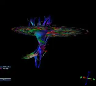
Diffusion Tensor Imaging or “DTI” is an MRI technique that enables the measurement of the restricted diffusion of water in tissue. It was first introduced to the field of MR by Peter Basser in 1994. It allows the mapping of water molecules in the brain and can be used to reconstruct the three dimensional white matter fiber tracts in the brain. DTI is particularly effective in the evaluation of the white matter of the central nervous system microstructure considered the most important region of abnormality in a Traumatic Brain Injury.
It is known that water diffuses asymmetrically in the white matter of the brain. In other words, water diffuses faster along the long axis (the length) of the axons. This property is known as anisotropy and can be used to define the direction of the axons. DTI takes advantage of diffusion anisotropy by measuring the anisotropy of the tracts to provide excellent details of the brain.
DTI has the ability to qualitatively and quantitatively demonstrate pathology that is not detected by other modalities such as CAT Scan and routine MRI. In other words, DTI has detected brain injury which a CT or normal MRI did not show. There is a consensus in the medical literature that DTI effectively differentiates patients with TBI from patients without it.
DTI has been documented as a powerful technique for in vivo detection of diffuse axonal brain injuries, also known as “DAI.” Diffuse axonal injury occurs when there is a force to the brain which causes shearing of the white matter. It is commonly seen at the junction of where the white matter and the gray matter of the brain meet. The force can cause the movement of the white matter in one direction and the movement of the gray matter in the opposite direction to produce a shearing or tearing of the white matter. Since this occurs at a microscopic level, it is undetected by a normal brain MRI or CT scan.