Heart attack is a leading cause of death and disability in the United States. There are major efforts underway to find improved methods for prevention, detection and treatment. These efforts are sponsored by many organizations including the American Heart Association, the American College of Cardiology, and the federal government.
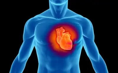
In order to best understand the science of heart attack, a basic knowledge of heart (cardiac) anatomy and function is necessary. The heart is basically a pump that receives blood lacking oxygen from the veins (venous system), pumps it to the lungs for oxygenation, and then returns it to the body thru the arteries (arterial system).
Anatomically the heart consists of 4 chambers whose walls are made of muscle. The right atrium initially receives unoxygenated blood from the venous system. Blood then enters the right ventricle, but contraction of the atrium helps pump more blood into the right ventricle. As the right ventricular chamber begins to contract, the tricuspid valve closes so that blood does not reenter the right atrium. Contraction of the right ventricle rejects blood into the pulmonary artery which is the major conduit carrying blood to the lungs.
As the right ventricle finishes its contraction, the pulmonic valve closes so that blood does not reenter the right ventricle from the pulmonary artery. Once blood is oxygenated in the lungs, it is returned via the pulmonary veins to the left atrium and left ventricle. When the left ventricle begins to contract, the mitral valve closes so that blood does not reenter the left atrium. The oxygenated blood from the left ventricle is pumped into the aorta which delivers blood via the many branches of the arterial system throughout the body. Once the left ventricle finishes its contraction, the aortic valve closes which prevents blood from reentering the heart.
There are microscopic and biochemical differences between heart muscle and regular muscle which is attached to skeletal bones. However, both types of muscle require their own nutrient blood supply through arteries. This nutrient blood supply provides oxygen and sugars without which contraction would not occur. The nutrient blood supply of the heart is supplied by the coronary arteries of which there are 2 and, in fact, they are the first branches off the aorta. They are located just beyond or distal to the aortic valve. The right coronary artery supplies the right ventricular chamber, and the left coronary artery supplies the left ventricular chamber.
The main portions of both arteries are 4 to 8mm. in diameter. Both arteries have many branches all of which decrease in size the farther they are from the main or proximal part. The atria receive blood from small branches of both of these arteries. It is the interruption of the coronary artery blood supply which causes a heart attack.
The bottom line determinant of whether or not a heart attack occurs is the oxygen and sugar (nutrient) demands of the heart muscle. One of these determinants is the thickness of the muscle making up the cardiac chamber walls. The atria are 2 to 3 mm. thick. The right ventricle is about 5 mm. thick, and the left ventricle is 10 to 12 mm. thick. The thicker the chamber wall, the more nutrient it requires, and, consequently, the left ventricular chamber is more vulnerable to heart attack. Another determinant is how often the heart contracts. A heart contracting at 150 time per minute uses substantially more oxygen and sugar than one contracting at 70 per minute.
Coronary artery occlusion is caused in the far majority of people by accumulation of cholesterol within the lumen and walls of the coronary arteries. The rate at which this cholesterol accumulates, the degree of blood supply interruption, and what happens to the cholesterol plaque itself in combination with nutrient demands determines what happens to the cardiac muscle.