The change from traditional open, large incision surgery to minimally invasive techniques is likely to be recorded as the most significant advance in the surgical art of the last part of the 20th century. Key to this trend is laparoscopy. Looking into the abdomen (the true meaning of the term laparoscopy), and operating with instruments through very small openings, allows surgery to be performed with far less postoperative discomfort and often faster recovery. Originally devised at the turn of the twentieth century, and utilized commonly by gynecologists before its widespread use in general surgery, the development of high-resolution microchip video cameras and specialized instruments has truly revolutionized intraabdominal surgery.
In order to visualize the organs and disease inside the abdomen, a space has to be created through which one can see the contents. This usually is accomplished by insufflating gas, usually carbon dioxide, and stretching forward the abdominal wall (similar to the stretching that occurs in pregnancy).
Instrumentation for laparoscopy:
• The laparoscope – a rod filled with fiberoptic fibers that transmit light and images without distortion. Sizes range from 10 mm in diameter down to 2 mm, and there are several different angles for viewing – most commonly a straight-on (O degree) ‘scope is used. The laparoscope has a lens on the outer end, through which the surgeon looked directly before the advances in video camera technology occurred. Today, a video camera is attached, and the surgeon and his assistants view the operation on high-resolution video monitors.
• A high intensity light source, transmitted by fiberoptic cable, to supply the light necessary to see inside the abdomen.
• An automatic insufflator – the device to pump (insufflate) gas into the abdomen under pressure and maintain steady distension of the abdominal wall so that a field of view is possible (pneumoperitoneum). By far, the most common insufflating gas is carbon dioxide. Its advantage is that it is inert and non-explosive, permitting cautery and other electrical equipment to be used safely inside the abdomen; its disadvantage is that it is irritating to the abdominal lining, and therefore general anesthesia is required for most laparoscopic procedures.
• The video camera, which either attaches to, or in some instances, is an integral part of, the laparoscope. The high resolution available with today's technology is phenomenal.
• A recording device, usually a printer of video images, to document important findings. Videotape sometimes also is used.
• Trochars: Different size tubes, commonly ranging from 2 to 12 mm in inner diameter, with valves on their outer ends, through which the instruments used during the procedure are inserted and removed, while maintaining the pneumoperioneum for visualization
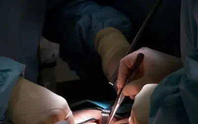
• Various instruments used by the surgeon – these are similar to instruments used in open surgery, only they are made to operate at the tips of long rods, permitting their introduction through the various sized trochars. They include scissors, graspers, retractors, cautery devices, stapling devices, clip appliers, needle holders and sutures, suction and irrigating systems, and other technology. In addition, instruments inserted into the uterus allow the organ to be manipulated, increasing access to the uterus, Fallopian tubes, and ovaries during gynecological procedures.
• Energy sources – most commonly electrocautery, occasionally lasers, and yes, even water, injected under pressure to separate tissues.
• The operating table itself, which by electrical manipulation can be moved into a variety of positions, helping the surgeon by allowing gravity to drop covering organs out of the way.
Basic Laparoscopic Techniques:
The first step in all laparoscopic procedures is accessing the abdomen -- allowing the insertion of the first trochar. This can be accomplished in either of two ways:
• The closed technique, in which a special retractable tip blunt ended needle (Veress needle) is inserted into the abdomen, usually through or just below the umbilicus (belly button), the gas is insufflated, and then an operating trochar is inserted after the abdomen is distended and a space created between the wall and the underlying organs.
• The open technique, in which a small incision is made, and the trochar tube introduced into the abdomen in a technique similar to that used in open surgery, only with a much smaller incision. Again this initial step usually is through or just below the umbilicus, where the abdominal wall is the thinnest.
Neither method has proven to be better than the other; many surgeons use both techniques, choosing which to utilize on a case by case basis.
By far the most dangerous step(s) are inserting the initial trochar (and/or Veress needle, if used). After all, one basically is trying to make a small “stab wound” into the abdomen without damaging any of the abdominal organs or blood vessels. Then, one or more additional operating trochars, of varying sizes, commonly are inserted. These secondary trochars are inserted while looking into the abdomen directly, making their placement inherently safer than the initial access.
Once the trochars are placed, the various instruments needed for a specific procedure are introduced through the trochars. Additional trochars may be placed during the course of a procedure as circumstances dictate. The opposing angles required to manipulate instruments at the ends of relatively long, thin tubes often results in the sites chosen for the small incisions used in laparoscopy being placed some distance away from the actual location of the disease.
Complications:
Risks are inherent in any surgical procedure, and laparoscopy is no exception. Each individual operation, such as cholecystectomy (gall bladder removal), appendectomy, tubal ligation, etc. has complications specifically related to the procedure. This section will deal with complications related to laparoscopy in general.
Perhaps the most worrisome problem is injury to an underlying organ or blood vessel while achieving access to the abdomen, most commonly while inserting the initial trochar and/or the Veress needle. Manufacturers have devised trochars with spring-loaded, retractable blades that cover the sharp tip almost instantly upon entering the abdomen, but injury is possible nonetheless. Most trochar injuries occur while inserting the first tube, which usually cannot be visualized upon entering the abdomen. Making the initial access away from sites of previous surgical procedures (scars), where adhesions are likely to be present and internal organs stuck to the abdominal lining, may reduce the chance of injury. Some surgeons feel that the open technique lessens the chance of injury; others feel the Veress needle is safe when used properly. The surgeon's experience and knowledge of his or her specific patient are most valuable. Some ingenuity in choosing the site for initial trochar placement may be necessary.
Complications can occur in establishing or maintaining the gas under pressure in the abdomen (pneumoperitoneum). If the abdominal pressure becomes too high, it may impair circulation or respiration. A dreaded complication of laparoscopy is the entrance of the gas into a vein, usually the result of inadvertently placing the Veress needle or a trochar directly into a major vessel. Starting the insufflation with a slow rate of flow until one is sure of proper placement helps avoid this problem.
Cautery and other energy sources are used during laparoscopy to cut tissues, stop bleeding, etc. Such energy sources can stray, and cause injury to organs away from the operative site. Often these injuries are not seen during the procedure, only becoming apparent later.
Laparoscopy allows only a two-dimensional view of the abdominal contents, and relies on the surgical team’s sense of vision. Depth perception is limited. One cannot usually feel the structures inside the abdomen and evaluate them directly.
Most laparoscopic procedures are well tolerated, quite safe, and many major operations that formerly required several days of hospitalization for recovery can now be done on an outpatient basis or with just a brief one or two day hospitalization. People should recover quickly and promptly after laparoscopic procedures. Complications are sometimes difficult to appreciate and evaluate. Patients and surgeons alike need be aware that if things do not seemingly “sail” smoothly after laparoscopy, there may be a hidden problem.
Prompt identification and treatment of the problem is essential. Excessive pain following laparoscopy is a significant “red flag” that a complication may be occurring. It is a grave mistake for a surgeon to disregard patient complaints after laparoscopy.