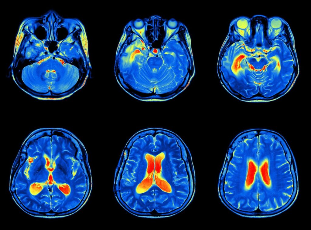
Neuroimaging is the process by which pictures or images of the brain are either directly or indirectly recorded onto a film and interpreted by a neuroradiologist, a radiologist who has additional specialty education and training in reviewing and interpreting these brain images.
Neuroimaging is classified into two broad categories: Structural Imaging and Functional Imaging. Structural imaging relates to the structures of the nervous system and the diagnosis of intracranial disease and injuries, including traumatic brain injuries. Functional imaging is used to diagnose metabolic diseases and lesions through visualization of the processing of information which will increase metabolism and “light up” on the scan.
There are many different types of brain imaging techniques:
- CAT Scan (CT);
- Magnetic Resonance Imaging (MRI);
- Functional Magnetic Resonance Imaging (fMRI)
- Positron Emission Tomography (PET);
- Single-Photon Emission Computed Tomography (SPECT);
- Magnetoencepholography (MEG);
- Diffuse Optical Imaging (DOI);
- Event-Related Optical Signal (EROS);
- Cranial Ultrasound.
Each of the above techniques have been developed to obtain visual information to aid in the diagnosis of brain pathology, including TBI.
In addition to these types of imaging techniques, there are subsets of techniques used to elicit further information from the type of scan being performed. For instance, diffusion tensor imaging is used in conjunction with a 3T MRI scan to evaluate how water molecules flow in the brain to help demonstrate diffuse axonal injury and other white matter abnormalities. NeuroQuant, is an FDA approved computer software that reviews 3T MRI scans to objectively measure hippocampal atrophy in patients who suffer TBI and who may have other conditions causing brain atrophy.