Single-Photon Emission Computed Tomography or SPECT, scan is a type of nuclear imaging study which measures blood perfusion or blood flow in tissues and organs. SPECT utilizes two imaging technologies; Computed Tomography and a radioactive tracer which allows physicians to see how blood flows. It is used in the brain to view how the blood flows in the arteries and vessels of the brain and can help diagnose brain injury in areas where there is diminished blood flow.
Although SPECT has been around since the 1950’s, it did not become widespread in clinical use until the 1980’s. SPECT scans can be used to analyze blood flow to many different tissues and organs, including the heart and brain.
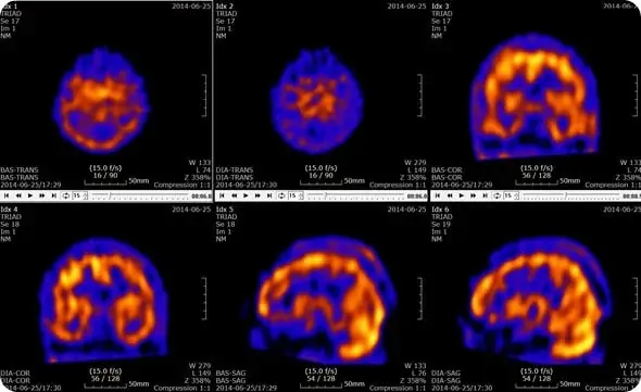
In the brain, a normal SPECT scan shows full, even, symmetrical perfusion. Common findings in TBI on SPECT include:
- focal decreased near sight of injury and/or opposite side (contra coup)
- asymmetrical hypoperfusion in the prefrontal, temporal, parietal or occipital lobes
- flattening of the prefrontal pole
- decreased anterior temporal poles
- decreased contralateral cerebellar perfusion.
SPECT aids in understanding TBI patients’ symptomatology and assists clinicians in developing targeted treatment strategies. SPECT helps evaluate perfusion abnormalities not only in blunt brain trauma, but also in cases of post-concussive syndrome and whiplash. Brain injured patients with normal EEG, CT, and/or MRI scans often complain of headaches, memory loss, concentration difficulties, dizziness, perceptual sensitivities, and emotional lability. Such patients may be labeled as malingering, when there are significant and demonstrable functional abnormalities present. Researchers investigating the differences between functional and structural imaging techniques have found SPECT to be more sensitive for patients with varying degrees of head trauma.