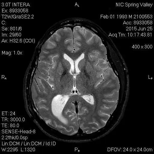
The human brain is fully encased in a bony vault called the skull. Inside the skull the brain is occupied by a clear fluid that suspends the brain within the skull for further protection. This clear fluid is called cerebrospinal fluid. Cerebrospinal fluid is produced in the ventricular system of the brain. The ventricles of the brain are a communicating network of cavities filed with cerebrospinal fluid and located within the brain that extend to form the central canal of the spinal cord. The cerebral ventricles consist of the lateral ventricles, third ventricle and fourth ventricle. The third ventricle is involved with protection of the brain from trauma and a pathway for the circulation of cerebrospinal fluid.
Peer-reviewed medical literature has determined that following a traumatic brain injury the ventricles of the brain can become damaged and changed. Dilated brain ventricles occur when the two halves of the brain are abnormally enlarged or swollen. A study of trauma patients that provided an assessment on average 30 years after TBI found that lateral ventricular enlargement was significantly associated with impaired memory functions, memory complaints and executive function. Of the MRI parameters used, the best predictor for cognitive outcome was the volume of the lateral ventricle. See for example Cognitive functions in relation to MRI findings 30 years after traumatic brain injury, Himanen L, Portin, R, Brain Inj. 2005 Bef. 19(2) 93-100.
Thus, MRI evaluation of the lateral ventricles is recommended in patients who have suffered a TBI as an important prognostic indicator of severity and outcome in TBI.