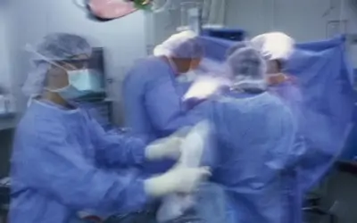
Thoracoscopic surgery of the chest was first described in terms of its original concept in 1922 by Dr. Jacobaeus. He was far ahead of his time in terms of originality of thought and almost 50 years ahead of the technology needed to make this exciting new diagnostic and therapeutic tool available for clinical use. In 1970, Dr. Joe Miller, Jr., at the Emory Clinic, began to match changes in technology with clinical applications and the field of thoracoscopic surgery was born. My first experience did not come until the early 1990ties when thoracoscopy emerged from lab use and was available for every day practice.
The thoracoscope consists of a slender fiberoptic tube than can be inserted into a 1/2 inch incision in the chest. The image is then combined with a tiny telescopic lens, a powerful light source, and a small video camera and is projected onto a TV screen. The surgeon can literally see into the chest.
Then using graspers, endoscopic scissors, and endostaples, the surgeon can perform a whole host of procedures. The revolutionary changes this technique has brought to thoracic surgery mark a milestone in the evolution of surgical technology. Thoracoscopy offers many patients marked advantages over standard open procedures. First, it gets the patient home from the hospital in 36 to 48 hours after the procedure. Second, recovery time from surgery and the level of pain experienced by the patient is markedly reduced. Lastly, the small incisions used are better tolerated than the old larger open thoracotomy incisions.
The most familiar use of thoracoscopy is to diagnose disease within the chest wall. In this case, small pinch biopsies of the pleura, the membrane surrounding the lung, the chest wall, the lung, and the pericardium surrounding the heart can be obtained. In addition, the mediastinum or, the area between the two lungs, can be readily visualized and lymph nodes biopsied. In patients with fluid collections around the lung called pleural effusions, samples can be obtained for culture studies and cells for cytological examination.
Patients with asbestos exposure who may have concerns about pleural tumors called mesotheliomas are usually good candidates for this technique. Other patients with abnormal chest x-rays and pulmonary nodules within the lung may benefit from small wedge resections made with the help of an endostapling device. Often, the lesions removed can be determined to be a benign tumor such as sarcoid or a malignant cancer of the lung. Using the wedge technique, small to moderate sized masses can be completely removed and sent to pathology for examination under the microscope.
A second common use for thoracoscopy is therapeutic. A number of topics fall into this grouping. Treatment of pleural disease, removal of pus collections call empyemas and lysis of adhesions from entrapped lungs are but a few indications. Other applications of this new technique involve removal of blebs or weak areas on the surface of the lung, staging of lung cancers and resections of metastatic disease from other areas of the body. In terms of the heart and the pericardium, pericardial effusions can be drained and cardiac tamponade relieved.
In my own practice I have operated on a series of patients with excessive sweating called hyperhidrosis, Raynaud's disease and reflex sympathetic dystrophy. In these cases, the sympathetic chain of nerves passing on the inside of the chest wall can be resected and sweating and pain reduced to symptomatic arms, hands and fingers. Another group of patients with esophageal conditions such as acid reflux disease and benign tumors of the esophageal wall can be helped.
This is but a quick review of a revolutionary new technique available to the thoracic surgeon to diagnose and treat a wide variety of conditions found in the chest. More indications seem to come along each year all to the patient's benefit.
Recommendations:
1) If you have an abnormal chest x-ray and are told that a mass, nodule, lymph node or fluid collection is present, a test called a CT scan of the chest is usually recommended to better define or isolate the abnormality.
2) A second test called a bronchoscopy may then be indicated. This test involves using a small tube with a light on the end to look down the airways in hopes of seeing the lesion and possibly getting a biopsy.
3) A third test called a CT directed needle biopsy may then aspirate cells from the mass or collect fluid samples.
4) Finally, you may be referred to a thoracic surgeon for the new thoracoscopy procedure. Hopefully, this single test may be used to both make the diagnosis and effect the removal of the abnormal area. If all else fails, the old open thoracotomy can be held in reserve to allow a hands on approach to the problem. Good Luck and I hope you benefit from the new fiberoptic technology.